Neurabin-II, also called spinophilin, interacts with actin and PP-1 in dendritic spines of the central nervous system (1,2). The gene encoding human neurabin-II maps to chromosome 17q21-q22 (2). The structural characteristics of neurabin-II include one F-actin binding domain at the N-terminal region, a predicted coiled-coil struture at the SLCterminal, one PDZ domain at the middle region, and a domain known to interact with transmembrane proteins (1). Neurabin-II bundles actin fliaments in vitro (1). In vivo, spinophilin localizes to the cortical sites of actin filaments and to the sites of active membrane remodelling (4). Neurabin-II also forms a complex with the catalytic subunit of PP1 and modulates PP1 enzymatic activity in vitro (2). Neurabin-II localizes to the head of dendritic spines (2) and aids in the ability of PP-1 to regulate the activity of a-amino-3-hydroxy-5-methyl-4-isoxazolepropionic acid (AMPA) and N-methyl-D-asparate (NMDA) receptors (3). In this manner, neurabin-II modulates both glutamatergic synaptic transmission and dendritic morphology (3). Synergistic interactions between spinophilin and human tumor supressor ARF suggest a role for neurabin-II in cell growth (5).
Function:
Seems to act as a scaffold protein in multiple signaling pathways. Modulates excitatory synaptic transmission and dendritic spine morphology. Binds to actin filaments (F-actin) and shows cross-linking activity. Binds along the sides of the F-actin. May play an important role in linking the actin cytoskeleton to the plasma membrane at the synaptic junction. Believed to target protein phosphatase 1/PP1 to dendritic spines, which are rich in F-actin, and regulates its specificity toward ion channels and other substrates, such as AMPA-type and NMDA-type glutamate receptors. Plays a role in regulation of G-protein coupled receptor signaling, including dopamine D2 receptors and alpha-adrenergic receptors. May establish a signaling complex for dopaminergic neurotransmission through D2 receptors by linking receptors downstream signaling molecules and the actin cytoskeleton. Binds to ADRA1B and RGS2 and mediates regulation of ADRA1B signaling. May confer to Rac signaling specificity by binding to both, RacGEFs and Rac effector proteins. Probably regulates p70 S6 kinase activity by forming a complex with TIAM1 (By similarity). Required for hepatocyte growth
Subunit:
Interacts with DCLK2 (By similarity). Possibly exists as a homodimer, homotrimer or a homotetramer. Interacts with F-actin, PPP1CA, neurabin-1, TGN38 and D(2) dopamine receptor. Interacts with RGS1, RGS2, RGS4, RGS19 and ADRA1B, ADRA2A, ADRA2B, ADRA2C, CDKN2A, PPP1R2, RASGFR1 and TIAM1. Interacts (via SLCterminus) with SPATA13 (via SLCterminal tail).
Subcellular Location:
Cytoplasm, cytoskeleton (By similarity). Nucleus (By similarity). Cell projection, dendritic spine (By similarity). Cell junction, synapse. Cell junction, adherens junction (By similarity). Cytoplasm. Cell membrane. Cell projection, lamellipodium. Cell projection, filopodium. Cell projection, ruffle membrane. Note=Enriched at synapse and cadherin-based cell-cell adhesion sites. In neurons, both cytosolic and membrane-associated, and highly enriched in the postsynaptic density apposed to exitatory synapses. Colocalizes with PPP1R2 at actin-rich adherens junctions in epithelial cells and in dendritic spines (By similarity). Accumulates in the lamellipodium, filopodium and ruffle membrane in response to hepatocyte growth factor (HGF) treatment.
Post-translational modifications:
Stimulation of D1 (but not D2) dopamine receptors induces Ser-94 phosphorylation. Dephosphorylation of Ser-94 is mediated mainly by PP1 and to a lesser extent by PP2A. Phosphorylation of spinophilin disrupts its association with F-actin, but does not affect its binding to PP1.
Similarity:
Contains 1 PDZ (DHR) domain.
SWISS:
Q96SB3
Gene ID:
84687
Database links:
Entrez Gene: 84687 Human
Entrez Gene: 217124 Mouse
Entrez Gene: 84686 Rat
Omim: 603325 Human
SwissProt: Q96SB3 Human
SwissProt: Q6R891 Mouse
SwissProt: O35274 Rat
Unigene: 514323 Human
Unigene: 229087 Mouse
Unigene: 476821 Mouse
Unigene: 6764 Rat
| Picture |
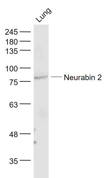
Sample:
Lung (Mouse) Lysate at 40 ug
Primary: Anti- Neurabin 2 (SL12146R) at 1/1000 dilution
Secondary: IRDye800CW Goat Anti-Rabbit IgG at 1/20000 dilution
Predicted band size: 89 kD
Observed band size: 89 kD
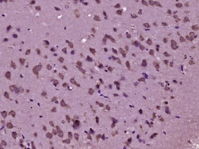
Paraformaldehyde-fixed, paraffin embedded (mouse brain tissue); Antigen retrieval by boiling in sodium citrate buffer (pH6.0) for 15min; Block endogenous peroxidase by 3% hydrogen peroxide for 20 minutes; Blocking buffer (normal goat serum) at 37°C for 30min; Antibody incubation with (Neurabin 2) Polyclonal Antibody, Unconjugated (SL12146R) at 1:400 overnight at 4°C, followed by operating according to SP Kit(Rabbit) (sp-0023) instructionsand DAB staining.
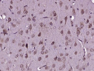
Paraformaldehyde-fixed, paraffin embedded (rat brain tissue); Antigen retrieval by boiling in sodium citrate buffer (pH6.0) for 15min; Block endogenous peroxidase by 3% hydrogen peroxide for 20 minutes; Blocking buffer (normal goat serum) at 37°C for 30min; Antibody incubation with (Neurabin 2) Polyclonal Antibody, Unconjugated (SL12146R) at 1:400 overnight at 4°C, followed by operating according to SP Kit(Rabbit) (sp-0023) instructionsand DAB staining.
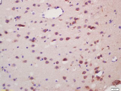
Tissue/cell: rat brain tissue; 4% Paraformaldehyde-fixed and paraffin-embedded;
Antigen retrieval: citrate buffer ( 0.01M, pH 6.0 ), Boiling bathing for 15min; Block endogenous peroxidase by 3% Hydrogen peroxide for 30min; Blocking buffer (normal goat serum,SLC0005) at 37℃ for 20 min;
Incubation: Anti-Neurabin 2 Polyclonal Antibody, Unconjugated(SL12146R) 1:200, overnight at 4°C, followed by conjugation to the secondary antibody(SP-0023) and DAB(SLC0010) staining
|
|
|